Beranda
/ Rib Cage Muscles And Tendons - Thoracic Wall And Breast Illustrations - If the force of the spasm is intense enough, muscle strains or tears in the tendons and ligaments can occur.
Rib Cage Muscles And Tendons - Thoracic Wall And Breast Illustrations - If the force of the spasm is intense enough, muscle strains or tears in the tendons and ligaments can occur.
Rib Cage Muscles And Tendons - Thoracic Wall And Breast Illustrations - If the force of the spasm is intense enough, muscle strains or tears in the tendons and ligaments can occur.. The human rib cage (thoracic cage) has the very important job of protecting the heart and lungs. They help stabilize your upper body and help you breathe. Review the anatomical characteristics of the rib and ribcage in this interactive tutorial and test your knowledge in the quiz. Tendons attach muscle to bone, muscles do not attach to bone. Quizlet is the easiest way to study, practise and master what you're learning.
Your intercostal muscles lie between your ribs. The ribs are part of the axial skeleton and are classified as flat bones. These muscles may be located anteriorly, posteriorly, and/or laterally. Less often, the lower legs, hands, and feet are painful and stiff. See more ideas about anatomy, rib cage anatomy, anatomy study.
Slipping Rib Syndrome Caring Medical Florida from www.caringmedical.com Any soft tissue (muscles, tendons, and ligaments) may be affected. Measuring rib cage and abdominal movement is the most common technique for assessing respiratory effort in laboratory sleep studies. Don't just draw a generic rib download assignment photos. This is a stereogram, to be viewed in crossview technique. But soft tissue of the neck, upper shoulders, chest, rib cage, lower back, thighs, arms, and areas around certain joints are especially likely to be painful. Introduction to the structure of the ribcage and ribs: Your intercostal muscles lie between your ribs, attaching them to one another. The joints in your rib.
A strain in this area can cause pain and difficulty breathing.
A strain in this area can cause pain and difficulty breathing. The underlying connective tissue may be temporarily or permanently damaged if. We look at the signs, causes, and treatment options here. Everyone has nice muscles in ct scanning! Construct a robo skelly rib cage and the pelvis using the bucket method. But soft tissue of the neck, upper shoulders, chest, rib cage, lower back, thighs, arms, and areas around certain joints are especially likely to be painful. Try to be as accurate as you can with them. The following general rules regarding actions can be. It provides a strong framework onto which the muscles of the shoulder girdle, chest, upper abdomen and back can attach. The sternum (breast bone) is made of 3 fused bones called the manubrium, body and the xiphoid process at the very tip. The intercostal muscles have different layers that are attached to the ribs to help build the chest wall and assist in breathing. Don't just draw a generic rib download assignment photos. Mesoderm, muscle, and tissue c.
Create your own flashcards or choose from millions created by a. The area under the ribs consists of intercostal muscle, ligaments and tendons, as well as the abdominal obliques, transverus abdominis and rectus abdominis just below the rib cage. This is a stereogram, to be viewed in crossview technique. Any soft tissue (muscles, tendons, and ligaments) may be affected. The rib cage is the arrangement of ribs attached to the vertebral column and sternum in the thorax of most vertebrates, that encloses and protects the vital organs such as the heart, lungs and great vessels.
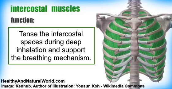
Intercostal Muscle Strain Causes Symptoms And Effective Treatments from www.healthyandnaturalworld.com Due to its cartilaginous attachments and surrounding tendons, the rib cage is. This is a large, triangular in shape, and flat muscle; The following general rules regarding actions can be. Beginning with one of the most dominant muscles in the region, the trapezius. Muscles that move the rib cage attach to the rib cage. It is formed by the 12 thoracic vertebrae, 12 pairs of ribs intercostal spaces are occupied by intercostal muscles and membranes, 11 intercostal nerves and two sets (main and collateral) of intercostal blood vessels also identified by. Intercostal muscles support respiratory function, while the upper abdominal muscles support your spine and. If the force of the spasm is intense enough, muscle strains or tears in the tendons and ligaments can occur.
In humans, the rib cage, also known as the thoracic cage.
They help stabilize your upper body and help you breathe. But soft tissue of the neck, upper shoulders, chest, rib cage, lower back, thighs, arms, and areas around certain joints are especially likely to be painful. Everyone has nice muscles in ct scanning! Your intercostal muscles lie between your ribs. The underlying connective tissue may be temporarily or permanently damaged if. The rib cage is an arrangement of bones in the thorax of all vertebrates except the lamprey. The rib cage, or thoracic basket, consists of the 12 thoracic (chest) vertebrae, the 24 ribs, and the breastbone , or sternum. Mesoderm, muscle, and tissue c. The thoracic cage (rib cage) is the skeleton of the thoracic wall. Muscles that position the pectoral girdle are all stated in great detail in the first table below, we will quickly explore some of them, their unique aspects, and their actions. We look at the signs, causes, and treatment options here. Often muscle spasms within the rib cage area are benign and caused by external forces such as injury. The sternum (breast bone) is made of 3 fused bones called the manubrium, body and the xiphoid process at the very tip.
The thoracic cage (rib cage) is the skeleton of the thoracic wall. Learn all about intercostal muscle strain, when the muscles between the ribs are damaged. Introduction to the structure of the ribcage and ribs: Measuring rib cage and abdominal movement is the most common technique for assessing respiratory effort in laboratory sleep studies. How to stretch out the muscles of the chest & rib cage.
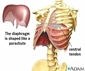
Vocal Technique Learn About The Diaphragm With Montreal Vocal Coach And Singing Teacher Lesley Findlay from vocaltechnique.ca Tissues, organs, and organ systems b. The underlying connective tissue may be temporarily or permanently damaged if. These muscles may be located anteriorly, posteriorly, and/or laterally. The intercostal muscles have different layers that are attached to the ribs to help build the chest wall and assist in breathing. Expiration (breathing out) is a result of relaxation of the respiratory muscles and of the elastic recoil of the lungs and of the fibrous ligaments and tendons attached to the. The rib cage, or thoracic basket, consists of the 12 thoracic (chest) vertebrae, the 24 ribs, and the breastbone , or sternum. It encloses and protects the heart and lungs. Don't just draw a generic rib download assignment photos.
It provides a strong framework onto which the muscles of the shoulder girdle, chest, upper abdomen and back can attach.
The rib cage has three important functions: How to stretch out the muscles of the chest & rib cage. Often muscle spasms within the rib cage area are benign and caused by external forces such as injury. Create your own flashcards or choose from millions created by a. Muscles that move the rib cage attach to the rib cage. Muscles that position the pectoral girdle are all stated in great detail in the first table below, we will quickly explore some of them, their unique aspects, and their actions. Volume rendering of a contrast enhanced thoracoabdominal ct scan. The intercostal muscles have different layers that are attached to the ribs to help build the chest wall and assist in breathing. There are twelve (12) pairs of ribs and all articulate posteriorly with the thoracic vertebrae. The thoracic cage, commonly called the rib cage, provides protection for the 2 lungs, heart, esophagus, diaphragm and liver. Quizlet is the easiest way to study, practise and master what you're learning. The ribs are part of the axial skeleton and are classified as flat bones. It is formed by the 12 thoracic vertebrae, 12 pairs of ribs intercostal spaces are occupied by intercostal muscles and membranes, 11 intercostal nerves and two sets (main and collateral) of intercostal blood vessels also identified by.
It provides a strong framework onto which the muscles of the shoulder girdle, chest, upper abdomen and back can attach rib cage muscles. Less often, the lower legs, hands, and feet are painful and stiff.
Berbagi
Posting Komentar
untuk "Rib Cage Muscles And Tendons - Thoracic Wall And Breast Illustrations - If the force of the spasm is intense enough, muscle strains or tears in the tendons and ligaments can occur."


Posting Komentar untuk "Rib Cage Muscles And Tendons - Thoracic Wall And Breast Illustrations - If the force of the spasm is intense enough, muscle strains or tears in the tendons and ligaments can occur."